
MRI
Magnetic resonance imaging is a medical imaging technique used in radiology to form pictures of the anatomy and the physiological processes of the body in both health and disease. MRI scanners use strong magnetic fields, electric field gradients, and radio waves to generate images of the organs in the body. MRI does not involve X-rays and the use of ionizing radiation, which distinguishes it from CT or CAT scans.
A head MRI is a useful tool for detecting a number of brain conditions, including:
- aneurysms, or bulging in the blood vessels of the brain
- multiple sclerosis
- spinal cord injuries
- hydrocephalus, a buildup of spinal fluid in the brain cavities
- stroke
- infections
- tumors
- cysts
- swelling
- hormonal disorders, such as acromegaly and Cushing’s syndrome
- hemorrhage, or bleeding
- inflammation
- problems with development or structure (such as a Chiari malformation)
- blood vessel issues
- an issue due to a previous head injury
A head MRI can help determine whether you sustained any damage from a stroke or head injury. Your doctor may also order a head MRI to investigate symptoms such as:
- dizziness
- weakness
- seizures
- changes in thinking or behavior
- blurry vision
- chronic headaches
These symptoms may be due to a brain issue, which an MRI scan can help detect.
A functional MRI (fMRI) of the brain is useful for people who might have to undergo brain surgery. An fMRI can pinpoint areas of the brain responsible for speech and language, and body movement. It does this by measuring metabolic changes that take place in your brain when you perform certain tasks. During this test, you may need to carry out small tasks, such as answering basic questions or tapping your thumb with your fingertips.
Additionally, there is a type of MRI called magnetic resonance angiography (MRA), which better examines the blood vessels in the brain.
The medical staff will need to know if you have any metal in your body, including:
- inner ear implants
- artificial joints
- a defibrillator or pacemaker
- particular types of heart valves
- vascular stents
- brain aneurysm clips
They’ll also ask whether you’ve ever worked with sheet metal or been injured with metal shrapnel. All of these things can affect how safely you can undergo an MRI. In the case of implants and pacemakers, those items can stop working properly due to an MRI’s powerful magnetic field.
If you’re wearing anything that contains metal, including jewelry or sunglasses, you will need to remove those items. Metal interferes with the MRI machine’s ability to produce a clear image. Braces and dental fillings typically won’t pose a problem, but pocketknives, pens, pins, and certain dental appliances can interfere. The staff may ask you to wear a hospital gown or clothing that doesn’t contain metal fasteners. You can’t have electronic devices in the MRI room.
Tell the medical staff if you’re pregnant. An MRI’s magnetic field affects unborn children in a way that isn’t yet fully understood.
Additionally, it’s important to let the staff know if you have claustrophobia. If so, you might need to take sedatives during the exam or have an “open” MRI. Open MRI machines have wider tunnels, which tend to be more tolerable for claustrophobic patients.
During the exam, it’s important to stay still to obtain the clearest images. Children who have difficulty staying still may need sedation, administered either orally or through an IV line. Sedation can also be helpful for adults who are claustrophobic.
You will lie down on a table that slides into the MRI machine. The table slides through a large magnet shaped like a tube. You may have a plastic coil placed around your head. After the table slides into the machine, a technician will take several pictures of your brain, each of which will take a few minutes. There will be a microphone in the machine that allows you to communicate with staff.
The test normally takes 30 to 60 minutes. You may receive a contrast solution, usually gadolinium, through an IV to allow the MRI machine to see certain parts of your brain more easily, particularly your blood vessels. The MRI scanner will make loud banging noises during the procedure. You may be offered earplugs to block the MRI machine’s noises, or you may listen to music during the test.
There are no risks associated with an MRI itself. There is a very slight chance that you will have an allergic reaction to a contrast solution. Tell the medical staff if you have decreased kidney function. It may not be safe to use contrast solution if this is the case.
A cervical spine MRI is usually used to diagnose the cause of neck pain. It’s often performed if the pain hasn’t improved with basic treatment. It may also be done if the pain is accompanied by numbness or weakness.
A cervical MRI scan can show:
- spinal birth defects or deformities
- an infection in or near the spine
- injury or trauma to the spine
- abnormal curvature of the spine, or scoliosis
- cancer or tumors of the spine
A cervical MRI may also be ordered before or after spinal surgery.
Ask your doctor if you can eat or drink before the scan, as protocols vary between facilities. Tell your doctor if you have diabetes or kidney problems if they want to use a contrast dye during the test. You may need a kidney function test before the scan. This will ensure that your kidneys can process the dye safely.
Tell your doctor if you’re pregnant. MRIs aren’t recommended during the first trimester of pregnancy. Your doctor may choose to postpone the scan until after you’ve had your baby.
Tell your doctor if you’re claustrophobic or have a fear of being in enclosed spaces. They can prescribe an antianxiety medication to help you feel more comfortable during the test. In some cases, you may be given anesthesia to put you to sleep.
Tell your doctor about any metal implants you have from a previous surgery. If so, it may not be safe for you to have an MRI scan.
Bring any relevant X-rays, CT scans, or previous MRI scans with you to your appointment. Sometimes the MRI technician will play music to help you relax. Bring a CD with you just in case.
Before you go in for the MRI, you’ll need to remove all jewelry and clothing that contains metal. It may be easier to leave your jewelry at home. You’ll probably need to wear a hospital gown during the test.
Your doctor might recommend an open MRI if you’re overweight or extremely claustrophobic. Open MRIs have slightly larger openings than standard machines. However, open MRIs aren’t available at all hospitals or clinics, so check with your doctor beforehand.
You’ll lie down on a narrow bed that’s attached to the MRI machine. Your head will be on a headrest and your arms at your sides.
The MRI technician will give you earplugs to muffle the loud knocking and thumping noises the machine makes when it’s running. You may have the option to listen to music during the scan. This can help relax you and distract you from the noise.
A frame called a “coil” will be placed over your head and neck. The coil contains an antenna. It helps focus the machine’s energy so it produces the most precise images. The MRI technician will also place a signaling device in your hand. You can use it to call for help while the test is running, if you need it.
Once you’re properly positioned, the table will slide into the machine. The MRI technician is able to see you through a window in an adjoining room. They’ll give you periodic updates on the scan’s progress.
Cervical MRI scans typically take 30 to 45 minutes. During this time, it’s very important that you stay as still as possible. The images may be blurred if you move.
What is MRI of the Chest?
Magnetic resonance imaging (MRI) is a noninvasive medical test that physicians use to diagnose medical conditions.
MRI uses a powerful magnetic field, radio frequency pulses and a computer to produce detailed pictures of organs, soft tissues, bone and virtually all other internal body structures. MRI does not use ionizing radiation (x-rays).
Detailed MR images allow physicians to evaluate various parts of the body and determine the presence of certain diseases. The images can then be examined on a computer monitor, transmitted electronically, printed or copied to a CD or uploaded to a digital cloud server.
MRI of the chest gives detailed pictures of structures within the chest cavity, including the mediastinum, chest wall, pleura, heart and vessels, from almost any angle. MRI also provides movie-like sequential imaging of the cardiovascular system that is important to assess the health and function of these structures (heart, valves, great vessels, etc.).
What are some common uses of the procedure?
MR imaging of the chest is performed to:
- assess abnormal masses, including cancer of the lungs or other tissues, which either cannot be assessed adequately with other imaging modalities (typically CT) or which are particularly well-suited to MR imaging.
- determine tumor size, extent, and the degree of spread to adjacent structures.
- assess the anatomy and function of the heart and its component structures (valves, etc.).
- assess myocardial perfusion (blood flow to the heart) and myocardial infarct (scar in the heart muscle due to prior obstruction of blood flow).
- determine blood flow dynamics in the vessels and heart chambers.
- display lymph nodes and blood vessels, including vascular and lymphatic malformations of the chest.
- assess disorders of the chest bones (vertebrae, ribs and sternum) and chest wall soft tissue (muscles and fat).
- assess for pericardial (thin sac around the heart) disease.
- characterize mediastinal or pleural lesions seen by other imaging modalities, such as chest x-ray or CT.
A special form of MRI called magnetic resonance angiography (MRA) is helpful to assess the vessels of the chest cavity (arteries and veins). MRA can also demonstrate an abnormal ballooning out of the wall of an artery (aneurysm) or a torn inner lining of an artery (dissection). See the MRA page for more information.
How should I prepare?
You may be asked to wear a gown during the exam or you may be allowed to wear your own clothing if it is loose-fitting and has no metal fasteners.
Guidelines about eating and drinking before an MRI exam vary with the specific exam and with the imaging facility. Unless you are told otherwise, you may follow your regular daily routine and take food and medications as usual.
Some MRI examinations may require you to receive an injection of contrast material into the bloodstream. The radiologist, technologist or a nurse may ask if you have allergies of any kind, such as an allergy to iodine or x-ray contrast material, drugs, food, or the environment, or if you have asthma. The contrast material most commonly used for an MRI exam contains a metal called gadolinium. Gadolinium can be used in patients with iodine contrast allergy. It is far less common for a patient to have an allergy to a gadolinium-based contrast agent used for MRI than the iodine-containing contrast for CT. However, even if it is known that the patient has an allergy to the gadolinium contrast, it may still be possible to use it after appropriate pre-medication. Patient consent will be requested in this instance. For more information on adverse reactions to gadolinium-based contrast agents, please consult the ACR Manual on Contrast Media.
You should also let the radiologist know if you have any serious health problems, or if you have had any recent surgeries. Some conditions, such as severe kidney disease, may prevent you from being given gadolinium contrast for an MRI. If you have a history of kidney disease or liver transplant, it will be necessary to perform a blood test to determine whether the kidneys are functioning adequately.
Women should always inform their physician or technologist if there is any possibility that they are pregnant. MRI has been used for scanning patients since the 1980s with no reports of any ill effects on pregnant women or their unborn babies. However, because the unborn baby will be in a strong magnetic field, pregnant women should not have this exam in the first three to four months of pregnancy unless the potential benefit from the MRI exam is assumed to outweigh the potential risks. Pregnant women should not receive injections of gadolinium contrast material except when absolutely necessary for medical treatment.
If you have claustrophobia (fear of enclosed spaces) or anxiety, you may want to ask your physician for a prescription for a mild sedative prior to your scheduled examination.
Infants and young children usually require sedation or anesthesia to complete an MRI exam without moving. Whether a child requires sedation depends on the child’s age, intellectual development and the type of exam. Moderate and conscious sedation can be provided at many facilities. A physician or nurse specializing in sedation or anesthesia for children should be available during the exam for your child’s safety. You will be given special instructions for how to prepare your child for the sedation or anesthesia.
Alternatively, certain pediatric facilities have child life personnel who can work with younger children to help avoid the need for sedation or anesthesia. They prepare the children for MRI by showing them a dummy scanner, play the noises that the child might hear during the MRI exam, answer any questions and explain the procedure to relieve their anxiety. Some pediatric facilities also provide goggles or headsets so that the child can watch a movie while the scan is being performed. Thus, the child remains motionless allowing for good quality images.
Jewelry and other accessories should be left at home, if possible, or removed prior to the MRI scan. Because they can interfere with the magnetic field of the MRI unit, metal and electronic items are not allowed in the exam room. In addition to affecting the MRI images, these objects can become projectiles within the MRI scanner room and may cause you and/or others nearby harm. These items include:
- jewelry, watches, credit cards and hearing aids, all of which can be damaged
- pins, hairpins, metal zippers and similar metallic items, which can distort MRI images
- removable dental work
- pens, pocket knives and eyeglasses
- body piercings
In most cases, an MRI exam is safe for patients with metal implants, except for a few types. People with the following implants cannot be scanned and should not enter the MRI scanning area:
- cochlear (ear) implant
- some types of clips used for brain aneurysms
- some types of metal coils placed within blood vessels
- nearly all cardiac defibrillators and pacemakers
You should tell the technologist if you have medical or electronic devices in your body. These objects may interfere with the exam or potentially pose a risk, depending on their nature and the strength of the MRI magnet. Many implanted devices will have a pamphlet explaining the MRI risks for that particular device. If you have the pamphlet, it is useful to bring that to the attention of the scheduler before the exam and bring it to your exam in case the radiologist or technologist has any questions. Some implanted devices require a short period of time after placement (usually six weeks) before being safe for MRI examinations. Examples include but are not limited to:
- artificial heart valves
- implanted drug infusion ports
- artificial limbs or metallic joint prostheses
- implanted nerve stimulators
- metal pins, screws, plates, stents or surgical staples
If there is any question of their presence, an x-ray may be taken to detect and identify any metal objects. In general, metal objects used in orthopedic surgery pose no risk during MRI. However, a recently placed artificial joint may require the use of another imaging procedure.
Patients who might have metal objects in certain parts of their bodies may also require an x-ray prior to an MRI. You should notify the technologist or radiologist of any shrapnel, bullets, or other pieces of metal that may be present in your body due to prior accidents. Foreign bodies near and especially lodged in the eyes are particularly important because they may move during the scan, possibly causing blindness. Dyes used in tattoos may contain iron and could heat up during an MRI scan, but this is rare. Tooth fillings and braces usually are not affected by the magnetic field, but they may distort images of the facial area or brain, so you should let the radiologist know about them.
Parents or family members who accompany patients into the scanning room also need to remove metal objects and notify the technologist of any medical or electronic devices they may have.
Abdominal MRI scans are used for a variety of reasons. Your doctor will order an MRI if they suspect something is wrong in your abdominal area but can’t determine what through a physical examination.
Your doctor may want you to undergo an abdominal MRI scan to:
- examine blood flow
- examine your blood vessels
- investigate the cause of pain or swelling
- examine lymph nodes
There have been no documented side effects from radio waves and magnetism to date.
Metal objects are not allowed near MRIs because the machine uses magnets. Let your doctor know if you have any metal implants, have worked in the metal industry, or have retained metal fragments from gunshot wounds, shrapnel, or another injury.
People who are claustrophobic or get nervous in enclosed spaces may feel uncomfortable in the machine. Your doctor may prescribe antianxiety medication or sedatives to help you relax.
Because the MRI uses magnets, it can attract metals. Alert your doctor if you have any type of metal implant from previous surgeries, such as:
- artificial heart valves
- clips, pins, or screws
- plates
- staples
- stents
Before the test, tell your doctor if you have a pacemaker. Depending on the type of pacemaker, your doctor may suggest a different radiological exam, such as an abdominal CT scan. Some pacemaker models can be reprogrammed before an MRI so they’re not disrupted during the examination.
If your doctor needs images of your colon, you may be required to use laxatives or enemas before the MRI. You also may need to fast for 4 to 6 hours before the exam.
Your doctor may require the use of a special dye that highlights areas of concern. This dye (gadolinium) is administered through an IV. While allergic reactions to the dye are rare, you should alert your doctor of any concerns before they give you the IV.
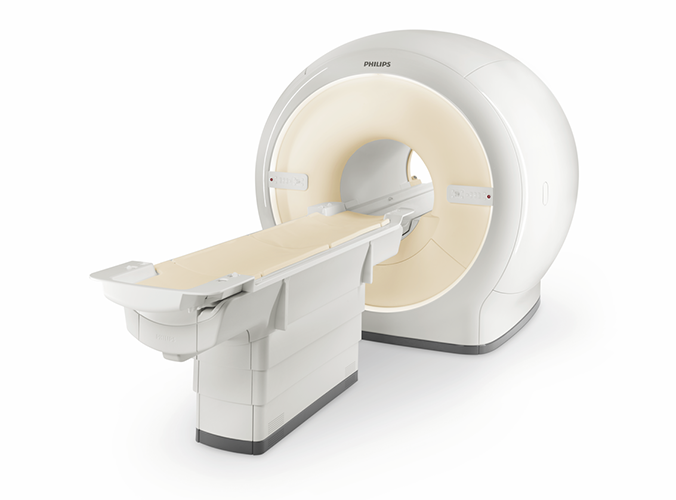
An MRI machine looks like it could transport you to another dimension. It has a bench that slowly glides you into a large tube attached to an opening that looks like a doughnut.
The technician will ask you to lie on your back on the bench and will give you a blanket or pillow. The technician will control the movement of the bench using a remote control from another room, and they will communicate with you over a microphone.
The machine will make loud whirring and thumping noises as it takes images. Many hospitals offer earplugs, televisions, or headphones to help pass the time.
MRI machines are very sensitive to movement, so it’s important that you stay very still. The technician may also ask you to hold your breath for a few seconds as pictures are being taken.
You won’t feel anything during the test. The magnets and radio frequencies are similar to those on FM radios, and can’t be felt.
The entire process takes 30 to 90 minutes.
Magnetic resonance imaging (MRI) of the prostate uses a powerful magnetic field, radio waves and a computer to produce detailed pictures of the structures within a man’s prostate gland. It is primarily used to evaluate the extent of prostate cancer and determine whether it has spread. It also may be used to help diagnose infection, benign prostatic hyperplasia (BPH) or congenital abnormalities. Exams may be performed using an endorectal coil – a thin wire covered with a latex balloon – that is inserted a short distance into the rectum. Prostate MRI does not use ionizing radiation, and it provides images that are clearer and more detailed than other imaging methods.
Tell your doctor about any health problems, recent surgeries or allergies. The magnetic field is not harmful, but it may cause some medical devices to malfunction. Most orthopedic implants pose no risk, but you should always tell the technologist if you have any devices or metal in your body. Guidelines about eating and drinking before your exam vary between facilities. Unless you are told otherwise, take your regular medications as usual. Leave jewelry at home and wear loose, comfortable clothing. You may be asked to wear a gown. If you have claustrophobia or anxiety, you may want to ask your doctor for a mild sedative prior to the exam.
What are some common uses of the procedure?
The primary indication for MRI of the prostate is the evaluation of prostate cancer. The test is commonly used to evaluate the extent of prostate cancer in order to determine if the cancer is confined to the prostate, or if it has spread outside of the prostate gland.
Occasionally, MRI of the prostate is used to evaluate other prostate problems, including:
- infection (prostatitis) or prostate abscess.
- an enlarged prostate, called benign prostatic hyperplasia (BPH).
- congenital abnormalities.
- complications after pelvic surgery.
How should I prepare?
Your MRI exam may, but will not necessarily involve the use of an endorectal coil, a thin wire covered with a latex balloon, placed inside the tail end of the large bowel, called the rectum. The rectum is located inside the pelvis immediately behind and up against the prostate gland. Placing this coil into the rectum so close to the prostate helps generate more detailed images from the prostate and surrounding structures; it also enables your radiologist to perform magnetic resonance (MR) spectroscopy, which can provide additional information on the chemical makeup of cells present in the prostate gland. Additionally, prostate MRI may examine water molecules motion (called water diffusion) and blood flow (called perfusion imaging) within the prostate to help differentiate abnormal (diseased) from normal prostate tissue.
You may be asked to wear a gown during the exam or you may be allowed to wear your own clothing if it is loose-fitting and has no metal fasteners.
Guidelines about eating and drinking before an MRI exam vary with the specific exam and with the imaging facility. Unless you are told otherwise, you may follow your regular daily routine and take food and medications as usual.
Some MRI examinations may require you to receive an injection of contrast material into the bloodstream. The radiologist, technologist or a nurse may ask if you have allergies of any kind, such as an allergy to iodine or x-ray contrast material, drugs, food, or the environment, or if you have asthma. The contrast material most commonly used for an MRI exam contains a metal called gadolinium. Gadolinium can be used in patients with iodine contrast allergy. It is far less common for a patient to have an allergy to a gadolinium-based contrast agent used for MRI than the iodine-containing contrast for CT. However, even if it is known that the patient has an allergy to the gadolinium contrast, it may still be possible to use it after appropriate pre-medication. Patient consent will be requested in this instance. For more information on adverse reactions to gadolinium-based contrast agents, please consult the ACR Manual on Contrast Media.
You should also let the radiologist know if you have any serious health problems, or if you have had any recent surgeries. Some conditions, such as severe kidney disease, may prevent you from being given gadolinium contrast for an MRI. If you have a history of kidney disease or liver transplant, it will be necessary to perform a blood test to determine whether the kidneys are functioning adequately.
If you are not allergic to latex, you may receive an endorectal MRI, otherwise you need to let the MR technologist know so that they may cover the endorectal coil with a latex-free condom. To prepare for an MRI with the endorectal coil, you should eat light meals on the day prior to and on the day of your exam. This will help make it easier to insert the coil. You may also be asked to use an enema preparation prior to your exam. An enema involves injecting liquid into your rectum through your anus to help clear the bowel. Enema kits or saline laxatives can be bought over-the-counter. Prior to your exam, you may continue to take your usual medications, unless you are told otherwise.
If you have claustrophobia (fear of enclosed spaces) or anxiety, you may want to ask your physician for a prescription for a mild sedative prior to your scheduled examination.
Jewelry and other accessories should be left at home, if possible, or removed prior to the MRI scan. Because they can interfere with the magnetic field of the MRI unit, metal and electronic items are not allowed in the exam room. In addition to affecting the MRI images, these objects can become projectiles within the MRI scanner room and may cause you and/or others nearby harm. These items include:
- jewelry, watches, credit cards and hearing aids, all of which can be damaged
- pins, hairpins, metal zippers and similar metallic items, which can distort MRI images
- removable dental work
- pens, pocket knives and eyeglasses
- body piercings
In most cases, an MRI exam is safe for patients with metal implants, except for a few types. People with the following implants cannot be scanned and should not enter the MRI scanning area:
- cochlear (ear) implant
- some types of clips used for brain aneurysms
- some types of metal coils placed within blood vessels
- nearly all cardiac defibrillators and pacemakers
You should tell the technologist if you have medical or electronic devices in your body. These objects may interfere with the exam or potentially pose a risk, depending on their nature and the strength of the MRI magnet. Many implanted devices will have a pamphlet explaining the MRI risks for that particular device. If you have the pamphlet, it is useful to bring that to the attention of the scheduler before the exam and bring it to your exam in case the radiologist or technologist has any questions. Some implanted devices require a short period of time after placement (usually six weeks) before being safe for MRI examinations. Examples include but are not limited to:
- artificial heart valves
- implanted drug infusion ports
- artificial limbs or metallic joint prostheses
- implanted nerve stimulators
- metal pins, screws, plates, stents or surgical staples
If there is any question of their presence, an x-ray may be taken to detect and identify any metal objects. In general, metal objects used in orthopedic surgery pose no risk during MRI. However, a recently placed artificial joint may require the use of another imaging procedure.
Patients who might have metal objects in certain parts of their bodies may also require an x-ray prior to an MRI. You should notify the technologist or radiologist of any shrapnel, bullets, or other pieces of metal that may be present in your body due to prior accidents. Foreign bodies near and especially lodged in the eyes are particularly important because they may move during the scan, possibly causing blindness. Dyes used in tattoos may contain iron and could heat up during an MRI scan, but this is rare. Tooth fillings and braces usually are not affected by the magnetic field, but they may distort images of the facial area or brain, so you should let the radiologist know about them.